Striatal neurones
The cell types of the striatum are well known (Kawaguchi, 1997), although not easy to identify in a brain slice. There are principal neurones (projection neurones), and interneurones as well. The projection neurones are all GABAergic, i.e., they use GABA as a neurotransmitter, and are the most abundant cell type. There are two kind of projection neurones; those that express Enkephalin (an endogenous opioid), and those that express substance-P, together with GABA. Morphologically, both classes are medium-size spiny neurones. It seems that they cannot be distinguished according to the morphology of the soma or dendrites.
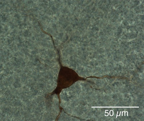
In the striatum, one finds both GABAergic and cholinergic interneurones. One class of GABAergic interneurones expresses the calcium-binding protein Parvalbumin. These cells are very similar to the hippocampal basket cells, and can fire action potentials at a very high frequency (~50 Hz). Sometimes, they have a small soma with a pear shape (pyriform). The other class of GABAergic interneurones express somatostatin, nitric oxide (NO), and neuropeptide Y. The cholinergic interneurones are easier to identify, but difficult to find. They have very large somata, sometimes 30 µm long, and often fusiform. They show a large sag in response to a hyperpolarising current, which is due to a hyperpolarising-activated cation current (Ih). This current is often found in cells that oscillate (the Ih current is also found in heart cells). Cholinergic interneurones comprise about 2% of striatal neurones.
|
(Picture by the author) |
posted by Alfredo @ 4:45 pm
![]()
![]()

0 Comments:
Post a Comment
<< Home